Wednesday, 21 March 2012

cDNA Libraries, Gel Electrophoresis and Dideoxy Sequencing

In this post we will be discussing some of the tools that are used by molecular biologists and biochemists to manipulate nucleic acids in order to diagnose and analyse disease. We'll discuss how cDNA libraries are constructed, how gel electrophoresis works as well as the basics of Dideoxy Sequencing. 

cDNA Libraries

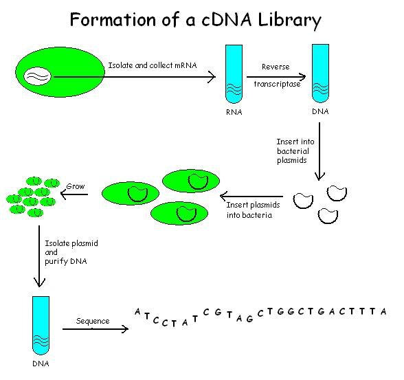
cDNA cloning is almost the same as genomic cloning (see previous post) except that the library only contains coding regions of the DNA (the structural part of genes). Genes may be cloned in order to understand the function of a gene, to mass produce a protein (eg. insulin) or to transfer a gene from one organism to another. Several steps are involved in the construction of a cDNA library:
  1. Isolate RNA
  2. Generate cDNA: To do this you have to:
      1. Isolate the mRNA from cells or tissue of interest using a mRNA Poly A tail
      2. Use dT primer and Reverse Transcriptase to synthesise a mRNA/DNA hybrid
      3. mRNA is degraded
      4. The free 3 end of the cDNA strand forms a hairpin loop that primes the synthesis of DNA using DNA Polymerase III.
      5. S1 nuclease cleaves the hairpin loop leaving a double stranded cDNA/DNA copy of the original mRNA
  3. Ligate into vector
  4. Introduce recombinant vector into E.coli
  5. Screen cDNA library for gene of interest: the library is plated out in a dish. Antibiotic is added in order to screen for the gene of interest. This is because cDNA has antibiotic resistance. 
  6. Analysis of cDNA clones.


The Formation of a cDNA Library
source:http://commons.wikimedia.org/wiki/File%3AFormation_of_a_cDNA_Library.jpg
If you'd like to use this file please see this website first
Gel Electrophoresis

In this technique, DNA molecules are placed in wells carved out of a section of agarose or polyacrylamide gel. A positive electrode is placed on one side of the gel while a negative electrode is placed on the other side. DNA molecules are negatively charged so they migrate to the positive pole. However, large molecules migrate slower than small molecules. DNA molecules of a known size are used to monitor the progression of the gel electrophoresis and to determine the size of the DNA molecules. Specific DNA fragments can be isolated from the gel and used for further analysis.

Gel Electrophoresis
source: http://commons.wikimedia.org/wiki/File%3AGel_electrophoresis_2.jpg
If you'd like to use this picture please see this website first.
 In the left hand column of the picture above, DNA molecules of known sizes were used as a reference. The lower the band is, the smaller the molecules are. The different intensities indicate different concentrations, the brighter the band the more DNA present.

Dideoxy Sequencing

This is used to sequence DNA. When DNA is being replicated, DNA polymerase requires a primer. The enzyme extends the primer by adding nucleotides to the strand of DNA. In this method, ddNTP's are added to the reaction. ddNTP's lack a 3' OH group and have a fluorescent marker. Because they lack a 3' OH group they cause the reaction to stop causing the chain of nucleotides to stop. The Polymerase Chain Reaction (PCR) is used to sequence the reaction. However only one primer is added (instead of 2 in normal PCR) and ddNTP's are added, so this process is termed Cycle Sequencing. The reaction will include:
  • The DNA template
  • TAQ polymerase
  • A primer
  • dNTP's
  • ddNTP's
 The reaction follows a cycle of:
  1. denaturation
  2. annealing
  3. extension
After many cycles have occurred there are multiple fragments of every length of DNA possible, each terminated by a ddNTP (dideoxy nucleotide). Unlike in PCR, however, there is a linear increase in the number of fragments and not a logarithmic increase.

Once this process is complete, the fragments are placed in a capillary tube and capillary electrophoresis is performed. An anode and cathode are placed at either end of the tube and the DNA migrate towards the anode. Smaller fragments of DNA migrate faster than larger fragments and a laser at the end of the tube detects the different wavelengths emitted by the ddNTP at the end of each fragment. This effectively reads the DNA sequence because the smaller fragments would have been the ones to have been terminated with a fluorescent marker first while the larger ones would have been terminated last. This data is then plotted as a chromatogram on a computer and the sequence can be analysed.  


PCR, Restriction Enzymes and Genomic Libraries

Hi :) Today's post will cover the next topic in our Veterinary Biochemistry unit - molecular tools. In this post I'll be discussing the Polymerase Chain Reaction (PCR) technique, restriction enzyme and how genomic libraries are constructed. 

Polymerase Chain Reaction (PCR):

The PCR technique is the amplification of nucleic acid sequences via repeated cycles of:
  1. denaturation,
  2. oligonucleotide primer annealing,
  3. and DNA polymerase extension using a heat stable polymerase. 
PCR generates billions of copies of target DNA or RNA. 
The components of a PCR reaction include: the DNA template; primers; dNTPs (nucleotides) including dATP, dTTP, dCTP, dGTP; and DNA polymerase (a heat stable DNA polymerase taken from thermophilic bacteria). 


The process is summarised really well in the video below:


Advantages of PCR:

The PCR reaction is a very specific assay, this is determined by the two primers used. PCR also allows you to start with very small amounts of DNA. This means that it can be used to analyse single cells or to amplify the starting material. PCR is not dependent on the DNA being intact, this is useful for forensic analysis. PCR only generates products when the binding sites for primers are present. This allows one to directly detect the presence of mutations because mutations prevent binding. This also allows PCR to be used as a diagnostic technique for the presence of foreign DNA. PCR can also be used in combination with other methods to amplify the amount of DNA available for analysis. 

Restriction Enzymes

Restriction enzymes are like DNA scissors. They recognise short DNA sequences and cut the DNA molecules at those specific sites. Their natural biological function is to protect bacteria by attacking viral and other foreign DNA. Some restriction enzymes make staggered cut that leave the resulting DNA with sticky ends. Enzymes which cleave the DNA at the centre of the recognition sequence leave blunt ended fragments of DNA. 

Genomic Libraries 


Genomic DNA libraries contain all the DNA of an organism. They are usually constructed for whole genome sequencing projects. There are 4 steps involved in constructing a DNA library:
  1. Extract and purify genomic DNA
  2. Digest DNA with restriction enzyme
  3. Insert into a suitable vector (eg. plasmid) and seal DNA with suitable ligase
  4. Plasmids containing foreign DNA are introduced (transformed) into bacteria and allowed to grow.
The plasmids which are in the bacteria give the bacteria resistance to antibiotics. This allows the particular bacteria to be isolated from other bacteria in a sample.