Hello :) The brain consists of several divisions including the: telencephalon, diencephalon, mesencephalon, metencephalon, and myencephalon. In this post, we'll take a look at each of these divisions.
When an animal embryo is developing, the brain starts out as a simple tube beneath the skin. As the embryo grows and develops, the anterior end of this tube swells and becomes the brain. Shortly after this, the brain forms three divisions which later expand and change to form 5 divisions, we'll go through each of these divisions below.
Forebrain
The forebrain is composed of the telencephalon and the diencephalon.
Telencephalon
An aside: The word 'telencephalon' is derived from the Greek word 'telos', meaning end, and the word 'encephalon' which refers to the brain. So telencephalon refers to the end of the brain. This may help you remember what part of the brain it refers to.
The telencephalon refers to the cerebrum, this includes the left and right cerebral hemispheres.The largest parts of the telencephalon are the left and right cerebral cortices which consist of the outer cortical grey matter. Below this grey matter is white matter which partly encloses the basal ganglia. The telencephalon contains the lateral ventricles (see this post). The telencephalon can be divided up into regions which are named after the bones of the cranium which cover them, these are shown in the diagram below. Blue represents the frontal lobe, green - temporal, yellow - parietal, and red - occipital.
 |
| Lobes of the Cerebrum (Telencephalon) |
Each of these areas contain specialised functional areas which are referred to as projection and association areas. The primary projection areas of the cerebral cortex are associated with certain somatic tracts which are projected via the thalamus. The thalamus is an area of the brain between the cerebral cortex and midbrain and is involved in relaying sensory and motor signals to the cortex. The projection areas are surrounded by a region called the association area which is involved in the processing and cognition of stimuli. The primary projection and association areas include:
- Somatic Motor Area: this is found in each of the cerebral hemispheres and functions in learnt motor skills. These are the origins of the cerebrospinal tract which is responsible for the initiation of the movements of the musculoskeletal system.
- Somatic Sensory: this is found in the parietal lobe and functions in conscious sensations. In regards to movement, it also senses tensions, positions of limbs and the forces acting on various body parts.
- Visual: located in the occipital lobe and involved in vision.
- Auditory: located in the temporal lobe and involved in hearing.
- Olfactory: this is located in a special area of the brain known as the piriform cortex, it functions in smell.
The telencephalon also includes the basal ganglia, which are also called basal nuclei. These are masses of grey matter which lie beneath the cortex within the white matter of the cerebral hemispheres. They are a series of discrete grey matter units that have many interconnections. Their job is to support the body's voluntary movements through postural adjustments as well as to assist or initiate the performance of automatic movements.
Neocortical Dominance
The neocortex is the outer part of the cerebral cortex and is involved in higher functions such as sensory perception and the generation of motor commands. Out of all the vertebrates, humans have the greatest ability to conceptualise, communicate, remember, associate and analyse input into their central nervous systems. The large neocortex of humans allows highly sophisticated and flexible responses to environmental change. Domestic animals also have many of these abilities but the extent varies between species.
Other species however, such as retiles and amphibians, have more stereotypical responses. Their responses to environmental stimuli are mainly reflexes. The more highly evolved the species is, the more it is able to vary its responses on the basis of learned and remembered past experiences.
Rhinencephalon
The telencephalon also includes an area of the brain known as the Rhinencephalon. This region is mainly related to olfaction and has olfactory and non-olfactory components. In the olfactory component, the axons of olfactory neuroepithelial cells pass through the cribiform plate and synapse with another neuron in the olfactory bulb. This neuron passes via interneurons to the cerebral cortex. The non-olfactory component includes the hippocampus which is an association area. This is believed to be involved with emotion, recent memory and autonomic function.
Limbic System
This includes the limbic lobe as well as the subcortical nuclei. The limbic lobe consists of the deep medial cortical grey matter while the subcortical nuclei includes structures such as the amygdala, hypothalamus, and hipocampus. This system receives and associates visceral, oral, sexual and basic sensory (olfaction, optic and auditory) impulses and then projects them to the hypothalamus. The system is also invloved with emotional and behavioural patterns via the hypothalamus through the autonomic nervous system.
Diencephalon:
Diancephalon refers to the most rostral part of the brain stem.
Another aside: The word 'diencephalon' is derived from the Greek word 'dia', meaning 'through' and, 'encephalon' which refers to the brain. So one way to remember where the diencephalon is located is to imagine looking through the brain :)
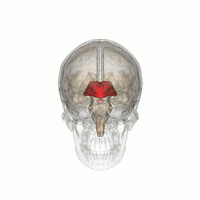 |
| The Diencephalon (red). Source. Please see this website if you'd like to use the animation. |
The diencephalon lies beneath the cerebral hemispheres, as shown in the animation above. In 'lower' vertebrates the diencephalon is the main association centre of the brain. However, in 'higher' vertebrates the cerebral hemispheres have taken over this role. The diencephalic nuclei still remain and are woven into the pathways that lead to and from the cerebrum. The largest part of the diencephalon is the thalamus, which I wrote a little about above.
The thalamus is a complex intercommunicating network which lies along the full length of the diencephalon on either side of the third ventricle. The two thalami are connected by an interthalamic adhesion. The thalamus is mostly made up of grey matter in the form of many closely packed thalmic nuclei which relate to specific pathways. The thalamus functions as:
- a relay centre for all sensory input to the cerebral cortex. This includes stimuli such as proprioception, pain and temperature, stimuli from the cerebellum as well as some stimuli from the basal ganglia.
- a recognition centre for some specific sensations such as tactile, thermal and pain sensations. In addition, there is a basic thalmic awareness of pain, touch, heat and vibration but there is poor localisation. Ie. the thalamus can tell something is going on but it can't tell where it is occurring.
The diencephalon also contains the hypothalamus which functions in regulating several hormones as well as other bodily functions such as eating, drinking and sleeping. The hypothalamus forms the floor of the brain and responds to neural input (this includes cortical input as well as ascending tracts from the brain stem and spinal cord). It also responds to factors within the circulating blood such as temperature, osmotic pressure and hormone levels.
The hypothalamus exerts is effects through the autonomic nervous system as well as the endocrine system. The anterior hypothalamus is the origin of the parasympathetic nervous system while the posterior is the origin of the sympathetic system.
The hypothalamus exerts is effects through the autonomic nervous system as well as the endocrine system. The anterior hypothalamus is the origin of the parasympathetic nervous system while the posterior is the origin of the sympathetic system.
Midbrain
The Mesencephalon
The mesencephalon refers to the midbrain.
Etymology: I find that knowing what the root of the word that we need to know is helps me to remember what it means. The word 'mesencephalon' is derived from the Greek word 'mesos', which means 'middle', as well as the word 'encephalon' which refers to the brain. So mesencephalon = 'middle brain'. Its easy to remember where the mesencephalon is if you know the origin of the word :)
The main role of the mesencephalon is as a channel for fibres to pass through. It also contains structures such as the red nuclei which are the origins of the rubrospinal tract. The mesencephalon has a stratified structure and is composed of several layers. The most dorsal of these layers is the tectum whose major features are four rounded surface swellings. The paired caudal swellings are known as the caudal colliculi and are the integration centres of auditory pathways. It receives auditory input from the inner ear and some of these impulses are transferred to the relevant part of the cerebral cortex for interpretation. Some impulses are also transferred to the rostral calliculus and ultimately to the tectospinal system in the reflex turning of the head towards the source of a sudden loud noise.
The rostral colliculi receive some input directly from the optic tracts. It also mediates reflex impulses such as blinking and pupillary adjustments.
The rostral colliculi receive some input directly from the optic tracts. It also mediates reflex impulses such as blinking and pupillary adjustments.
Hindbrain
Metencephalon
The main part of the metencephalon is the cerebellum but it also contains the pons. The main function of the cerebellum is to maintain motor synergy throughout the body by the coordination of motor activity, the maintenance of muscle tone and equilibrium. If an animal wants to move, the somatic motor area of the cerebral cortex will communicate its intentions to the cerebellum through corticopontine tracts which will ensure that these intentions are followed out. The cerebellum does this through pre-control, that is, the cerebellum compares the current state to the intended state and modifies the intended movement accordingly. The cerebellum also exerts feedback control to ensure that the movements occur smoothly. In addition, the cerebellum can remember information related to motor events, this is how people can learn to walk.
In terms of structure, the metencephalon lies above the fourth ventricle in the caudal cerebral fossa. It is connected to the brainstem by three pairs of peduncles, which are thick bundles of fibres. These are the superior, middle and inferior cerebellar peduncles. The cerebellum also has hemispheres which are connected to each other by the 'median vermis'. The cerebellum contains several important subcortical nuclei, these are:
- The dentate nucleus: this is the largest and most important and receives information from the cerebellar hemispheres. Its main role is limb coordination, posture and muscle tone.
- The fastigial nucleus: this receives information mainly from the vestibular apparatus and is mainly involved with equilibrium, muscle tone and axial posture.
The Myelencephalon
The myelencephalon refers to the medulla oblongata (or medulla for short). The medulla is located directly beneath the cerebellum and is physiologically important because of its many control centres and ascending and descending pathways. The control centres include those involved with respiration, cardiac and vasomotor activity as well as those regulating vomiting, swallowing, coughing, gastric secretion, urinating and defecating. In addition, it has the origin of the sixth to twelfth cranial nerves. The medulla is the most primitive region of the brain and functions mainly at a reflex level.
That's it for this post, if you have any questions please feel free to ask :)
I'm sad you didn't include the etymology for the metencephalon and the myelencephalon :(
ReplyDeleteOtherwise, great article! It's been super helpful.
Very nice summary and reading/teaching resource.
ReplyDeleteYes -- I'd like the etymology of the last two brain sections too -- in fact "myelencephalon -- word origin" was what I'd come looking for. But great post, regardless.
Very nice summary and reading/teaching resource.
ReplyDeleteYes -- I'd like the etymology of the last two brain sections too -- in fact "myelencephalon -- word origin" was what I'd come looking for. But great post, regardless.
Hi,
ReplyDeletenice article,
my doubt is ---> myencephalon and Myelencephalon are the same?
Hi, that was a great post. I really liked it. I learned a lot. I’m going to tell my friends about it. Thanks!
ReplyDeletehttps://blog.mindvalley.com/diencephalon-function/