Hi :) This post will discuss the ways in which the central nervous system (CNS) is protected, that is, by bone, meninges and cerebrospinal fluid. I'll discuss the bones and their relevant landmarks of the skull and vertebral column as well as the meninges of the CNS and the ventricles of the brain.
Bones
The Skull
The brain is encased by the skull which provides protection. This website has some good diagrams of what the skull looks like. I'm not going to go into too much detail here, the best way to learn these bones is to actually look at a real skull. I'd suggest going to an anatomy museum or lab, if you have access to one, to handle a skull. This is probably one of the best ways to learn the bones. You may also find it helpful to try and sketch and label the bones or to even quiz yourself with a game such as this. Another good way to remember the major bones of the skull is to use a mnemonic. One of the best I've heard (and probably the strangest) is: "I Never Masticate Lean Protein From Zebra Babies, Please Tell Our Moms".
- I (incisive),
- Never (Nasal),
- Masticate (Maxilla),
- Lean (Lacrimal),
- Protein (Palatine),
- From (Frontal),
- Zebra (Zygomatic),
- Babies (Basisphenoid wing),
- Please (Parietal),
- Tell (Temporal),
- Our (Occipital),
- Moms (Mandible)
The stranger the better because you're more likely to remember it!
The Vertebral Column
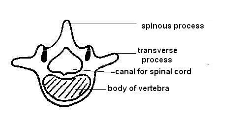
In dogs, the vertebral column is divided into 5 main regions: cervical (which includes 7 bones), thoracic (13), lumbar (7), sacral (3) and caudal (20). The general structure of a vertebra is shown below, although there are differences in the vertebra of different regions in the spinal cord. C1 and C2, the atlas and axis are heavily modified.
 |
| Source. Please see this website if you'd like to use the diagram. |
Meninges
Meninges are three continuous membranes which surround and help protect the brain and spinal cord.
Dura Mater
This is the tough outermost membrane and in the skull is fused with the inner periosteum.
Spinal Dura
The spinal cord only has a single layer of dura which is separated from the walls of the vertebrae by an epidural space to form a tube (called the dural tube). The epidural space is filled with fat and the internal vertebral venous plexus, which are veins inside the vertebral canal. The fat and veins cushion the spinal cord and allow it to adjust to the movements of the back and neck. The dural tube is attached to the upper surface of the caudal vertebrae. Distally, it tapers with the pia mater and anchors to the periosteum of the proximal coccygeal vertebrae to form the filum terminale. The role of this is to prevent the spinal cord from sliding cranially and caudally when the animal bends and moves around.
Cranial Dura
Because the dura mater is fused with the inner periosteum of the skull, no epidural space exists here. In the skull, where is it known as cranial dura, it is firmly attached to several sites within the cranial vault. This dura has two layers: the outer endosteal and inner meningeal. The cranial dura has a ventrally directed fold which lies longitudinally between the cerebral hemispheres named the falx cerebri. The dorsal sagittal venous sinus is present on the dorsal surface of the falx cerebri between its endosteal and meningeal layers. The caudal region of the falx cerebri attaches to the tentorium cerebelli.
The tentorium cerebelli is a part of the cranial dura which separates the cerebellum from the caudal end of the cerebral hemispheres. The core part of the tentorium is made up of bone and is surrounded by meninges.
Additionally, the cranial dura extends along the special sensory nerves. The dura runs extra cranially along the optic nerve until it inserts onto the sclera of the eye. The dura also follows the olfactory tract until the olfactory nerves are through the cribiform plate. The vestibulocochlear nerve is also surrounded by dura until it reaches the inner ear.
Arachnoid and Pia Mater:
The arachnoid and pia mater membranes are relatively delicate compared to the dura mater. The arachnoid lines the deep surface of the dura mater while the pia closely attached to and supports the brain and spinal cord, it also contains numerous small blood vessels. The arachnoid and pia mater are separated by the subarachnoid space which contains cerebrospinal fluid (CSF). The role of the subarachnoid space is to provide hydraulic protection to the brain and spinal cord, to act as a temporary reservoir for CSF, it also acts as a mechanism to alter cranial capacity and provides a route for blood vessels.
The inner surface of the arachnoid is joined to the pia by many trabeculae and filaments (this looks kind of like a spider's web, which is where the name subarachnoid comes from). The subarachnoid space contains cerebrospinal fluid.
The inner surface of the arachnoid is joined to the pia by many trabeculae and filaments (this looks kind of like a spider's web, which is where the name subarachnoid comes from). The subarachnoid space contains cerebrospinal fluid.
 |
Cerebrospinl Fluid
The cerebrospinal fluid (CSF) acts as a hydraulic skeleton within and surrounding the brain and works to buoy up and protect the brain and spinal cord. In addition, it protects the brain through chemical buffering and transports nutrients and removes wastes to and from the CNS. CSF is produced by the ependymal cells of the ventricles and central canal, the choroid plexuses and through selective leakage of vessels in the pia mater. Choroid plexuses are leaky tufts of arterioles, pia and ependymal epithelium within the brain's ventricles. Most of the CSF is produced by the choroid plexuses through dialysis from the arterioles and secretion from the ependymal cells.
CSF flows from the lateral ventricles through the interventricular foramina to the third ventricle then through the mesencephalic aqueduct to the fourth ventricle. Most of the CSF exits from each side of the lateral foramina into the subarachnoid space of the cerebellomedullary cistern. From there it flows to the surrounding brain and spinal cord.
Since CSF is derived from blood it must return to the blood stream after draining from the central nervous system. Within the central nervous system, the major drainage route for CSF are the arachnoid villi (aka. arachnoid granules). These villi are projections of the arachnoid membrane into the venous sinuses of the dura mater (such as the superior sagittal sinus). At each villus, CSF is separated from blood by flattened fibroblasts and endothelial cells. The villi act as valves which regulate the flow of CSF into the venous sinuses. When the pressure of the CSF is greater than venous pressure, the villi expand and the spaces between the cells increase, allowing more fluid to flow into the venous sinus. When the venous pressure is greater than the pressure of the CSF the villi collapse and this blocks the flow of blood into the subarachnoid space.
In addition, CSF can be absorbed directly by veins in the pia and the subarachnoid space due to an osmotic pressure effect.
Ventricles
These are an interconnecting series of cavities filled with CSF within the brain. There are four ventricles: the paired lateral ventricles as well as the third and fourth ventricles. There is one lateral ventricle within each cerebral hemisphere. The third ventricle is a narrow vertical space between the two lateral ventricles. An interventricular foramen allows each lateral ventricle to communicate with the third ventricle which is linked to the fourth ventricle by the mesencephalic aqueduct. The fourth ventricle lies beneath the cerebellum and above the pons and medulla. On the lateral sides of the fourth ventricle are lateral apertures which connects the ventricle to the subarachnoid space.
It's difficult to find diagrams showing the canine ventricular system but it should look something like this :)
It's difficult to find diagrams showing the canine ventricular system but it should look something like this :)
 |
| The Ventricular System |
That's it for this post, please feel free to leave any questions in the comments section below :)
No comments:
Post a Comment